Engineering tools to process many cells—and target individual ones
Last updated: October 13, 2026
Drug discovery and cell-based medicine need technologies that handle large populations and work with selected cells individually. We combine tiny channels and pores, light, microscopy and AI to pursue both speed and precision.
Our two priorities: high throughput × selective manipulation
01 Our research at a glance
How our research fits together
Process many cells while respecting individual differences
↓
DIRECTION AMany at onceHigh-throughput processingMicropores, microfluidics and parallel gel patterns
DIRECTION BChosen targetsSelective manipulationImaging, selected-cell encapsulation and laser intervention
The directions are complementary research aims, not mandatory consecutive experimental steps.
Delivering molecules, encapsulating cells and measuring cell responses are distinct techniques connected by one aim: precise manipulation and quantitative measurement at single-cell resolution.
02 Direction A | High-throughput parallel processing
Handling one cell at a time does not scale easily. We therefore arrange tiny structures in arrays to manipulate many cells in parallel.
A-1 Deliver molecules by squeezing cells through tiny pores
A cell membrane regulates what can enter. Pushing cells through 8–15 µm through-holes (1 µm = one-thousandth of a millimeter) temporarily increases membrane permeability, allowing research molecules and genetic cargo to enter.
Delivery through tiny holes
Up to ≈62,000 holes in the actual deviceOnly a small part of the array is illustrated
1. At the hole
Cells meet molecules in solution
2. Cell deforms
↓
Mechanical squeezing temporarily permeabilizes the membrane
3. Molecules enter
•••
Molecules diffuse into the cell
Green dots represent molecules. Schematic; not drawn to scale.
Up to 62,000 holes / 3 million cells per minute
Maximum reported in a collaborative 2025 study; delivery and viability depend on cell type and operating conditions.
A-2 Manipulate cells using arrays of micronozzles
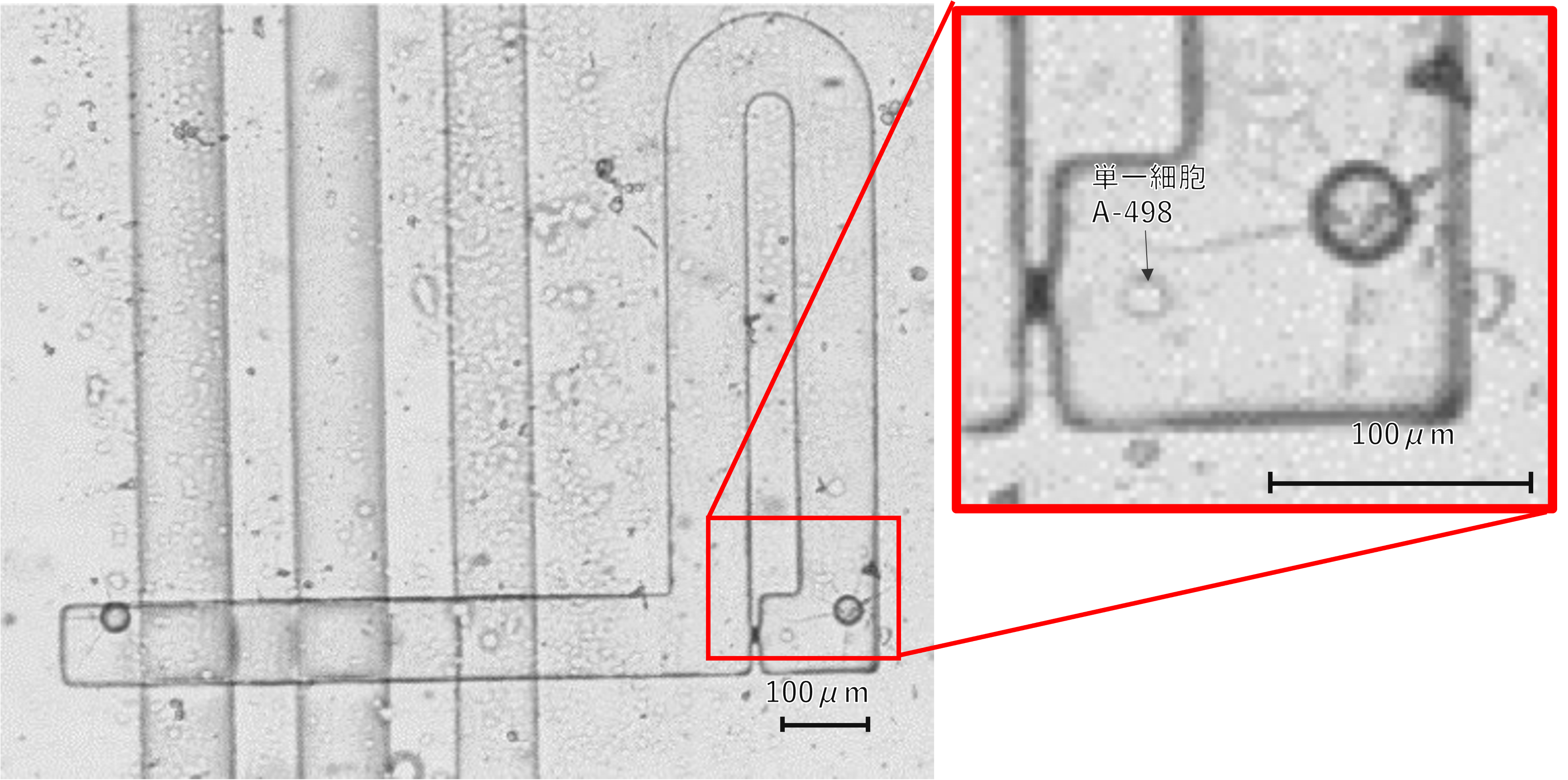
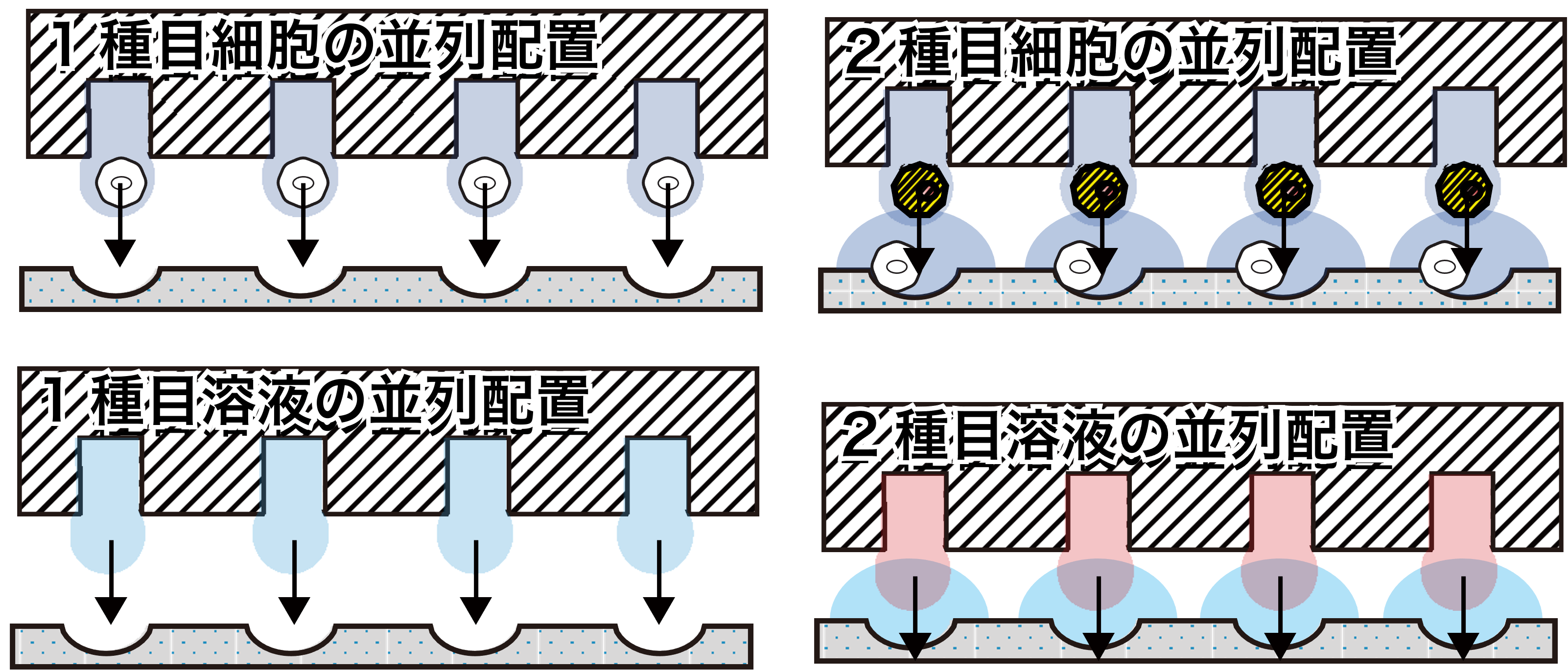
Micronozzles and nanoliter (nL) pipette arrays enable small-volume liquid handling and cell capture or positioning at multiple sites. We have also studied electrokinetic pumping for precise microscale actuation.
Many micronozzles act in parallel
↓ ↓ ↓ ↓ ↓
Microfluidic actuation, including electrokinetic flow, helps manipulate cells at multiple positions.
A-3 Pattern numerous small cell-containing hydrogels
Hydrogels are soft, water-rich materials. Using photocurable materials such as GelMA, we produce arrays of small gel compartments containing one or a few cells, supporting 3D culture. Nanohydroxyapatite (nHA) additives have also been studied for osteogenic differentiation and bone regeneration research.
Create many tiny hydrogel compartments
An array of hydrogel micropatterns can hold one cell or several cells, depending on conditions.
Pattern sizes: 80–250 µm
Collaborative 2025 study. The ≈97.2% viability result was reported for NIH-3T3 cells under particular conditions, not all cell types.
03 Direction B | Selective, precise cell manipulation
Individual cells vary in appearance and function. We investigate ways to find a particular cell in an image and apply an intervention at its location.
B-1 Identify a chosen cell and encapsulate it in a gel
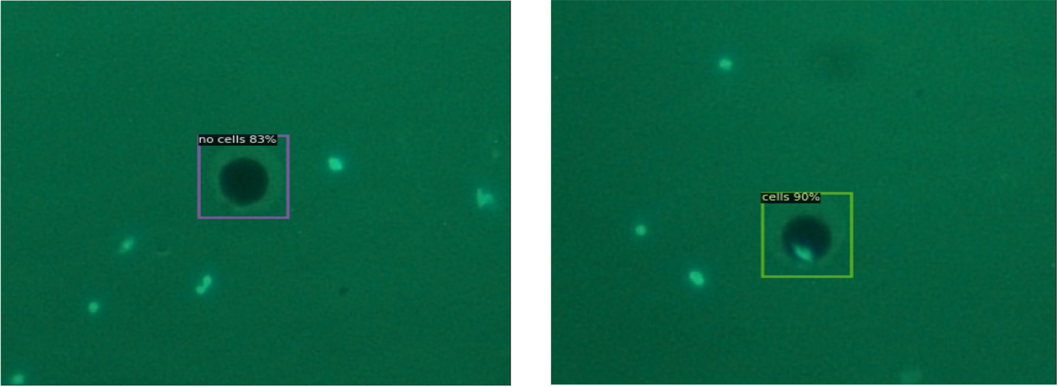
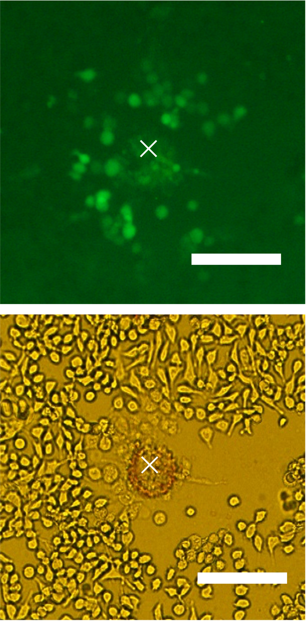
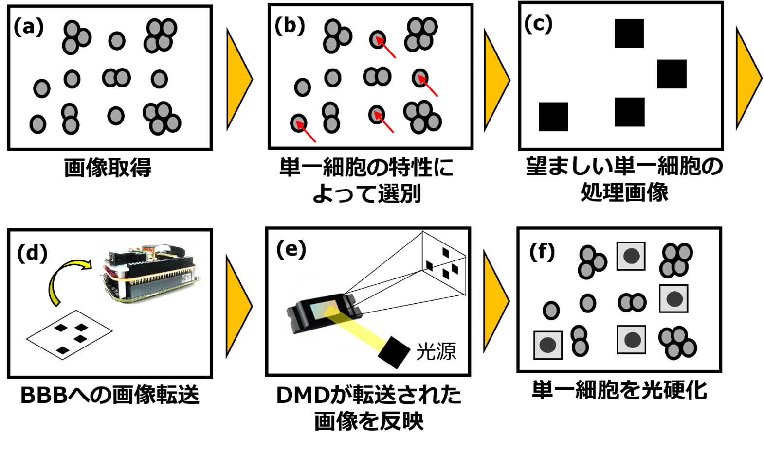
Microscope images identify cells and patterned light cures a local hydrogel around a selected target. Our 2025 conference contribution reported YOLOv8 detection of HeLa cells and selective photocuring in PEGDA. Related work is also exploring GelMA.
See cells, choose a target, cure gel around it
1. Observe
Locate cells in a microscope image
2. Select a target
Image analysis identifies a chosen cell
3. Cure hydrogel with light
Patterned illumination solidifies the local gel
A selection box marks the target and green shading shows the cured gel. The 2025 conference paper used HeLa cells and PEGDA.
B-2 Use laser-induced shockwaves for molecular delivery
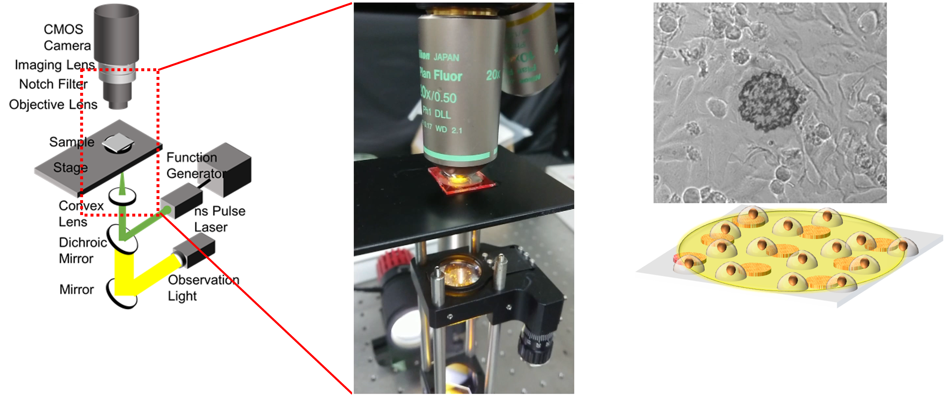
A short laser pulse irradiates a light-absorbing microdisk, creating localized mechanical effects that temporarily permeabilize cell membranes. We study how disk diameter, distance from cells and cell type change delivery and damage.
Use a laser-induced mechanical effect to permeabilize cells
1. Microdisk absorbs light
Microdisks are positioned near cells
2. A laser pulse creates a shockwave
Laser energy triggers a local mechanical effect
3. Nearby cells take up molecules
•••
Measure delivery and cell damage
Simplified from the 2025 microdisk-assisted optoporation study; the laser targets an absorbing microdisk.
2025 study: up to 60% delivery yield under optimized conditions
The study compared 20 µm and 50 µm disks. Delivery performance must be balanced against cell damage.
Parallel gel patterning (A-3) and selective encapsulation (B-1) serve different purposes. Selective encapsulation and optoporation are also separate methods, not mandatory consecutive steps.
04 Shared platform | Build, measure, improve
Both directions depend on fabrication, quantitative measurement, and data-driven improvement.
1. Build | Microfluidics and 3D printing
We design and fabricate tiny channels, microvalves, SlipChips and micronozzles using PDMS processes, masked stereolithography and direct ink writing (DIW), then verify device dimensions and flow performance.
3D-print designed microchannels
▼ ▼ ▼
Design, exposure conditions, fabricated channel dimensions and flow performance are evaluated.
2. Measure | Imaging and time-series analysis
Microscopy and image cytometry identify individual cells and quantify shape and fluorescence. Label-free time-lapse imaging can track cell responses over time, including drug-related effects.
Measure individual cells from images
Cell detection enables cell-by-cell measurements of morphology, fluorescence and viability. Bars are illustrative, not experimental data.
3. Improve | AI and Bayesian optimization
We investigate LLM-assisted CAD workflows and Bayesian optimization to improve geometries and 3D printing settings using experimental measurements.
CAD↔TEST↔AI
The design–experiment–improvement cycle
DesignDefine the goal and measurement
FabricateBuild and test the device
ManipulateProcess or select cells
EvaluateFeed measurements back into design
↶ Measurements inform the next design
This is not a claim that the entire workflow already runs autonomously. Published results and work under development are being combined toward more capable experimental workflows.
05 Related direction | Controlling Euglena with light
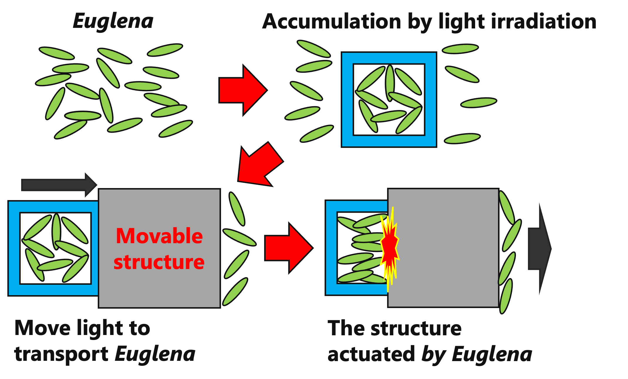
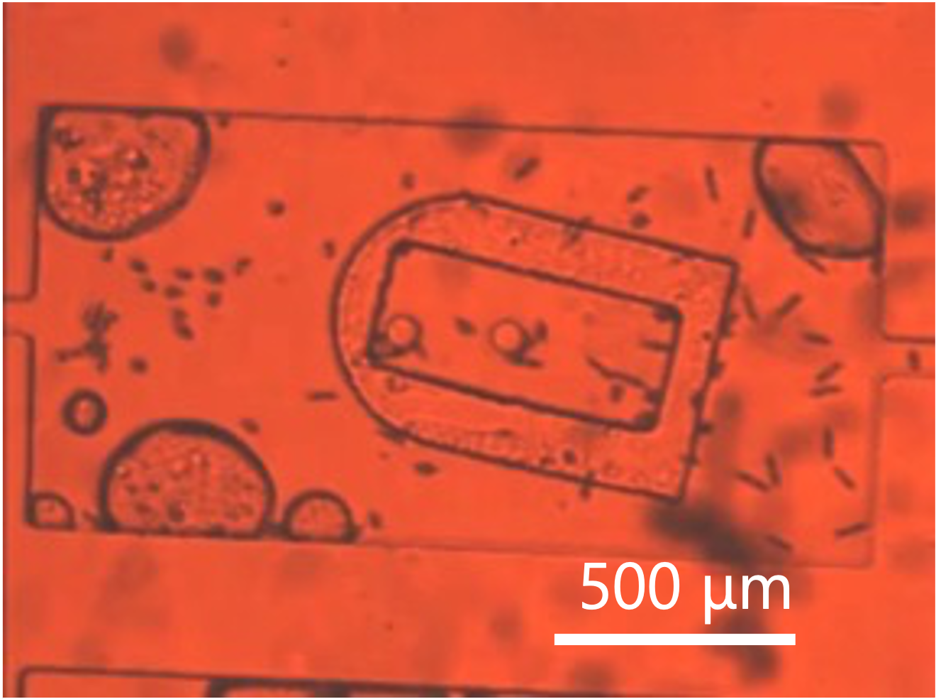
Alongside engineering cells as targets of manipulation, we explore biohybrid systems that use living cells as functional components. Euglena gracilis responds to light and swims through microscopic channels. Our research controls illumination to guide these movements.
Guide light-responsive Euglena in microchannels
☼
→
This work studies light-directed microorganism movement. Drug transport and oxygen delivery are possible future applications.
2025 study: light-directed ≈1 mm displacement (30–190 s)
Times depend on irradiation conditions. Drug transport and photosynthesis-based oxygen delivery are future application concepts, not demonstrated clinical treatments.
06 Research publications and presentations
These original explanatory diagrams simplify the underlying science. Refer to the journal papers and conference proceedings for actual microscopy images, operating conditions, and measurements.